By Harvey Society
Read Online or Download The Harvey Lectures: Series 102, 2006-2007 (Harvey Lectures Series) PDF
Similar history_1 books
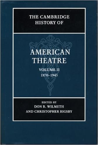
The Cambridge History of American Theatre: Volume 2: 1870-1945
Quantity starts off within the post-Civil struggle interval and lines the improvement of yank theater as much as 1945. It discusses the position of vaudeville, eu impacts, the increase of the Little Theater stream, altering audiences, modernism, the Federal Theater flow, significant actors and the increase of the megastar process, and the achievements of amazing playwrights.
- The Sindh-Punjab Water Dispute 1859-2002
- Jamaican Rock Stars, 1823-1971: The Geologists Who Explored Jamaica (Geological Society of America Memoir 205)
- Ernest Trumpp and W.H. McLeod as scholars of Sikh History, religion, and culture
- The 2000-2005 World Outlook for Elementary and Secondary Schools (Strategic Planning Series)
- The English Novel In History 1840-95 (Novel in History)
- Holocene Palaeoenvironmental History of the Central Sahara: Palaeoecology of Africa Vol. 29, An International Yearbook of Landscape Evolution and Palaeoenvironments ... of Africa and the Surrounding Islands)
Extra info for The Harvey Lectures: Series 102, 2006-2007 (Harvey Lectures Series)
Example text
It is interesting to remember that AGG2 and AGG3 both have paralogs. , 2005). A fourth paralog (155892) was not found in these studies and may not be a flagellar protein. Both the AGG2 and AGG3 paralogs are likely to have arisen by a recent duplication as the paralogs are tightly linked to each other. It is interesting to speculate that different pairs of paralogs may be found in the cis and trans flagella to help impart different responses to light signals. Tagged versions of the paralogs will be needed to pursue this hypothesis.
4. 5. c Fig. 1. (a) Electron micrograph of a Chlamydomonas cell preserved by cryofixation methods to illustrate the position of the pair of basal bodies (indicated by *) in the cell at the anterior end. ) (b) Diagram of basal body and transition zone structures. The distance from the base to the tip is approximately 450 nm. ) Ring of amorphous material. ) Triplet microtubules, also known as microtubule blades, with pinwheel. ) The A, B, and C tubules of one triplet microtubule are indicated. The central shaft is devoid of the pinwheel, but γ-tubulin has been localized to this region.
24 ROLE OF BASAL BODIES IN GENERATING ASYMMETRY a b 2 1 c 5 3 4 Fig. 3. The fiber systems attached to the Chlamydomonas basal body complex. (a) The mature basal bodies are shown in red, the transition zones in peach, and the probasal bodies in pink. The microtubular rootlets have four microtubules (orange) or two microtubules (yellow) and attach at specific triplet microtubules of the basal body. The distal (solid) and proximal (striped) striated fibers are shown in light blue (here, the latter are partially hidden by the former).


